


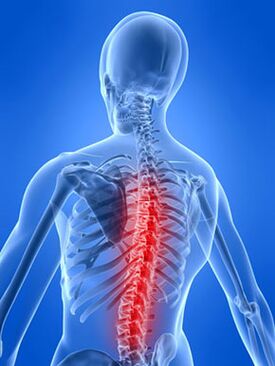
Thoracic osteochondrosisis a dystrophic degenerative change in the intervertebral discs of the thoracic spine.
This section of the spine consists of 12 vertebrae. It is the least mobile and is well protected by a muscular corset. Due to these properties, thoracic osteochondrosis is a rarer pathology than osteochondrosis of the cervical or lumbar spine. But, given the general trend towards an increase in the incidence of osteochondrosis, cases of osteochondrosis localized in the thoracic region are becoming more and more common.
Causes of thoracic osteochondrosis
The main cause of thoracic osteochondrosis, like other types of osteochondrosis, is degenerative changes in tissues and deterioration of metabolic processes due to malnutrition and irrational load on the intervertebral discs. Thoracic osteochondrosis most often occurs as a result of prolonged sitting in an irrational and uncomfortable position - at an office desk, while driving a car, as well as in the presence of scoliosis, which creates an uneven load on the spine. The nature of the pain that occurs with thoracic osteochondrosis determines two types of symptoms of this disease - dorsago and dorsalgia.
The manifestation of dorsago is expressed by acute intense pain, which has the nature of a sudden attack. In addition to limited back mobility, breathing difficulties may occur.
On the contrary, with dorsalgia, pain localized in the area of the affected discs is long-lasting, relatively mild and accompanied by limited mobility in the lumbar-thoracic or cervicothoracic spine.
The spinal canal in the thoracic region is quite narrow. Therefore, even with small-sized protrusions and hernias in thoracic osteochondrosis, compression of the spinal cord can occur. This condition is especially dangerous becausemay cause problems with the heart, liver, kidneys and pancreas. Therefore, timely treatment of thoracic osteochondrosis is so important to prevent complications.
The peculiarity of thoracic osteochondrosis is that its symptoms can easily be mistaken for signs of other diseases. Therefore, this disease is called the "chameleon disease. "In addition to cardiovascular diseases such as angina and heart attack, thoracic osteochondrosis imitates pain from appendicitis, cholecystitis, renal colic, as well as from diseases of the gastrointestinal tract such as gastritis, peptic ulcer, colitis (gastralgic syndrome).
If thoracic osteochondrosis is suspected, a thorough diagnosis must be carried out in order to be able to differentiate thoracic osteochondrosis from other diseases.
Symptoms of thoracic osteochondrosis
With thoracic osteochondrosis, a feeling of pain and discomfort appears. It is observed in the heart, chest, back, side, and upper abdomen. The pain intensifies with inhalation and exhalation, and with movement. Numbness of the left arm and interscapular area may be felt, which may require an ECG. With thoracic osteochondrosis, pain similar to intercostal neuralgia may occur, which radiates to the scapula.
Often, pain caused by thoracic osteochondrosis worsens at night, as in a heart attack, causing fear of death, and therefore can be mistaken for heart pain with suspected angina pectoris. Their difference from angina attacks is that pain during thoracic osteochondrosis is not relieved by nitrates, and the ECG does not reveal pathological signs characteristic of this disease. At the same time, taking heart medications is absolutely ineffective; pain relief is achieved by treating the disease itself.
If the symptoms of thoracic osteochondrosis depend on the location and mechanisms that caused the pathological process, most often the disease is accompanied by compression of the spinal roots. A much less common complication of thoracic osteochondrosis is compression of the spinal cord.
Symptoms of compression of radicular structures (radiculopathy)
Often thoracic osteochondrosis is manifested by radiculopathy, which develops when a herniated intervertebral disc appears. It can occur at any level, but hernias of the more mobile lower segment are most common. Symptoms of radiculopathy appear immediately after physical activity and slowly increase over several weeks.
If the symptoms and clinical manifestations of thoracic osteochondrosis are associated with a protrusion or herniation of a disc located in the upper segment of the thoracic spine, this will be pain in the shoulder, shoulder joint, scapula, chest or abdominal cavity.
Basically, the symptoms of thoracic osteochondrosis depend on the direction of the hernia: it is lateral or median. Thoracic osteochondrosis, which is complicated by a protrusion or lateral hernia, will be accompanied by unilateral pain, in addition, local loss of sensitivity and pain at the level of the hernia may appear. When a lateral hernia occurs, the symptoms of compression are minimal and reversible. The pain will intensify with movements of the spine, coughing, or taking a deep breath. When a median hernia occurs, the pain is prolonged and persistent, it can last for weeks. The main danger in this case may be due to compression of the spinal cord structures.
Thoracic osteochondrosis and compression of the spinal cord (compressive myelopathy)
Myelopathy of the thoracic spine can be quite rare. This is due to functional and anatomical features. In this case, the symptoms of thoracic osteochondrosis are local or surrounding pain, numbness, weakness in the legs, and dysfunction of the pelvic organs. The pain can radiate to the groin, abdomen, intercostal space or spread to the legs.
Clinical manifestations of thoracic osteochondrosis
Thoracic osteochondrosis as an independent disease or in combination with osteochondrosis of other parts of the spine is common. However, the clinical manifestations of this type of osteochondrosis, compared with osteochondrosis of the cervical and lumbar spine, are observed much less frequently, and the detected syndromes are not clearly expressed.
Thoracic osteochondrosis is not clinically manifested by "looseness" of the SDS or displacement of adjacent vertebrae. Arthrosis processes in the upper and lower segments of the thoracic spine, which in structure and function are close to the lower cervical and upper lumbar segments, on the one hand, are characterized by corresponding syndromes and symptoms for cervical and lumbar osteochondrosis. At the same time, they manifest characteristic clinical signs inherent only in the thoracic spine. These include intercostal neuralgia, costovertebral and costotransverse arthrosis, which are manifested by pain of varying intensity, intensifying with deep inspiration and coughing. Often constant, less often paroxysmal. With intercostal neuralgia, pain points are determined along the intercostal spaces. With costovertebral and costotransverse arthrosis, the pain intensifies with pressure on the ribs and is localized in the area of the paravertebral line.
Vertebrogenic syndromesat the thoracic level - primarily reflex manifestations: muscular-tonic, neurodystrophic and vasomotor. Difficult to differentiate vertebrogenic muscular-tonic, dystrophic and vascular reflex manifestations of the thoracic level, accompanied by pain in the back, are defined as dorsalgia, and in the area of the anterior chest wall - as pectalgia, if a more specific syndrome cannot be established.
Thoracic osteochondrosis, along with static and neurological disorders, is characterized by reflex visceral disorders of the heart, gastrointestinal tract, and genitourinary system. Pain in the heart area (pseudoanginal syndrome) can occur as a reflex response to irritation of the receptors of the affected cervical and upper thoracic spine. Vertebrogenic pseudoanginal pain differs from anginal pain not only in location, but also in the duration of attacks, in their dependence on the position of the spine, and in the ineffectiveness of nitrates. These are the so-called pectalgia, oranterior chest wall syndrome. Anterior chest wall syndrome should be considered in three variants, caused by cervical, thoracic and cervicothoracic pathology. With all these options, painful and reflex muscular-tonic, dystrophic and neurovascular changes develop in the pectoralis major muscle and other tissues of the anterior chest wall. The pain intensifies with physical exertion on the muscles of the chest, when turning the head and torso, but not after emotional, general physical stress or eating, like angina pain.
Compression syndrome, which occurs as a result of prolapse of a larger posterior herniated disc in the thoracic spine, is quite rare. At the same time, compression of the root is manifested by girdle pain and hypalgesia in the corresponding dermatome, and compression or compressive ischemia of the spinal cord (myelopathy) resembles the symptoms of an extramedullary tumor: pain, hypoalgic, motor and pelvic spinal disorders.
In most cases, with thoracic osteochondrosis, the disease develops rather slowly and initially manifests itself only in minor pain, localized in the back and intensifying after prolonged static load or other stay in one position. Over time, the intensity of pain increases and appears even with a short static load, and neurological symptoms often occur. In advanced stages of thoracic osteochondrosis, the pain becomes excruciating and does not depend on the position of the body and even intensifies at night.
Treatment of thoracic osteochondrosis
To treat thoracic osteochondrosis, reflex treatment methods can be used. To restore back mobility and eliminate spasms and muscle hypertonicity, acupuncture or, as they say in English-speaking countries, acupuncture is used. The use of this effective method allows you to improve the functioning of blood vessels, which has a beneficial effect on the nutrition and blood supply to the tissues of the intervertebral discs. The effectiveness of acupuncture used for thoracic osteochondrosis can be significantly increased through combined use with manual therapy, vacuum therapy, physiotherapy, cupping massage, and moxotherapy. These methods demonstrate high efficiency and safety, and therefore form the basis for a treatment course that is prescribed to patients individually. Using these methods, it is possible to stop the progression of the disease, return the intervertebral discs to normal functions, stimulate tissue regeneration (the fibrous ring of the disc and the nucleus pulposus), completely eliminate unpleasant symptoms of the disease, such as pain, and also prevent complications of osteochondrosis, which can manifest ashernias and protrusions.
In case of thoracic osteochondrosis, therapeutic exercises are of no small importance, which not only complements the main therapy, but also helps to form the correct muscle corset, thereby preventing future relapses.