


Currently, arthrosis is one of the most common pathologies of the musculoskeletal system and most often occurs in people aged 40 to 60 years. Until quite recently, this disease mainly affected pensioners, but now the situation is changing due to well-known reasons - a sedentary lifestyle, irregular diet and injuries contribute to the development of degenerative processes in the joints even in relatively young people.
According to forecasts, in the coming years the number of patients with deforming arthrosis will only increase; already their total number is about 8%. DOA of the shoulder joint and other joints is one of the main causes of loss of performance and disability.
Causes and mechanism of development
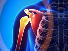
Shoulder arthrosis is a chronic pathology that primarily affects the cartilaginous tissues covering the articular surfaces of the bones. However, this does not mean that the cause is disturbances in the cartilage itself: arthrosis is a multifactorial disease and develops under the influence of a number of external circumstances.
Deforming arthrosis of the shoulder joint is called omarthrosis and can also affect the acromioclavicular joint (the junction of the shoulder blade and collarbone). There are several main reasons contributing to the occurrence of the disease:
- high physical activity associated with excess weight and sports training;
- injuries, congenital and acquired skeletal anomalies - kyphosis, scoliosis, varus or valgus deformity of the lower extremities, as well as improper fusion of bones after fractures;
- impairment of the regenerative ability of cartilage due to inflammatory, hormonal disorders or insufficient blood circulation;
- accelerated wear of intra-articular elements due to lack of joint fluid.
The shoulder joint is the most mobile because it forms a ball-and-socket joint. This is the freest joint in which movement can occur around many axes. Despite the fact that in practice a person uses only 3 axes of rotation, the shoulder is often subject to various dislocations and subluxations. That is why the most common is traumatic arthrosis of the shoulder joint.
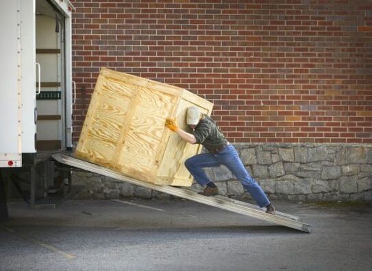
The group at increased risk of developing post-traumatic arthrosis includes men who have crossed the age limit of 60 years. Most of the patients are people who work in heavy production (loaders, builders) and athletes. Damage occurs due to frequent and sudden changes in pressure between the bones within the joint.
Since the right hand is dominant in most people, arthrosis of the right shoulder joint is most often diagnosed.
The following factors can provoke shoulder arthrosis:
- surgical interventions on the joint;
- genetic predisposition;
- intoxication with toxic substances at home or at work;
- hormonal changes in the postmenopausal period in women;
- hypothermia;
- disorders of a neurodystrophic nature in the cervical or lumbar segment of the spine (humeral periarthritis, iliopsoas muscle syndrome).
The immediate cause of dystrophic changes in the joint is a decrease in the ability of cartilage to self-heal. Normally, cartilage tissue is smooth, elastic and strong. During the development of arthrosis, it gradually loses its properties, becomes rough and exfoliates. As a result, chips appear on the cartilage, which "float" in the joint cavity and injure the synovial membrane.
The progression of the disease leads to calcification, ossification and the appearance of cysts in the cartilage tissue, as well as thickening of the joint capsule and inner membrane. Due to the thinning of the cartilage, the bones are practically exposed and begin to deform, and bone spines - osteophytes - form along the edges.
An increase in load on the muscular-ligamentous apparatus provokes fibrous degeneration of tissues and susceptibility to various sprains and tears. Sometimes the joint can "go" into a state of subluxation. In advanced stages, motor ability sharply decreases, and bone ankylosis develops (fusion of the articular surfaces of bones).
Stages and symptoms
Deforming arthrosis of the shoulder joint develops unnoticed and in most cases makes itself felt unexpectedly. Since there are no blood vessels and nerve endings in the cartilage, the first symptoms appear only when the pathological process has gone beyond the joint.
Pain is the most characteristic sign of arthrosis, and pain is clearly related to physical activity and weather conditions. When the shoulder is affected, pressing and aching pains occur, as well as dull and aching pains that radiate to the forearm and hand. The pain prevents you from moving your shoulder or arm, so your range of motion is significantly reduced.
Symptoms of arthrosis of the shoulder joint are:
- pain that intensifies when raising or moving the arm back;
- the lower edge of the collarbone or shoulder blade is painful and hot to the touch;
- the shoulder looks swollen and red;
- stiffness and crunching when moving.
Attention:Sometimes it is difficult to understand what exactly hurts - the elbow, the hand or the whole arm. Therefore, timely diagnosis is very important to determine the causes of pain.
Shoulder arthrosis develops in three stages, with its symptoms becoming more intense. At first, only discomfort and slight pain are felt after prolonged physical activity. In a state of rest, everything passes without a trace.
At the first stage of arthrosis, damage to the cartilage tissue is insignificant, but on x-rays you can see some narrowing of the joint space, the outlines of which change from round to elongated.
The second stage persistently announces itself with persistent pain, which does not always go away even at rest. Stiffness and limited movement increases; it is most difficult to move the arm back. At this stage, patients most often seek medical help, since manifestations of arthrosis significantly reduce the quality of life.
The situation is aggravated by the fact that, due to pain, a person avoids unnecessary movements. This leads to weakening and subsequent atrophy of the muscles surrounding the joint. Radiological signs of arthrosis of the second stage are joint deformations, bone growths and narrowing of the interarticular space.
Attention:in the second stage, arthrosis is much more treatable than in the third, when only surgery can help.
When moving to the third stage, the pain becomes unbearable and haunts the person constantly. In order to somehow alleviate the condition, you have to take a certain position. The pain syndrome no longer depends on movements, and the upper part of the arm loses the ability to perform any activity.
The final stage of shoulder arthrosis is the fusion of bones in the joint - bone ankylosis, in which the shoulder stops moving at all.
Diagnostics
The diagnosis of shoulder oarthrosis is made based on visual signs and radiographic results. It is worth noting that the severity of clinical symptoms does not always correspond to what the x-ray shows. However, some patterns still exist, so there are several diagnostic criteria:
- Stage 1– the joint space may remain the same or narrow slightly, osteophytes are necessarily present;
- Stage 2– the interarticular space is narrowed, pronounced bone growths are observed, bone deformations are possible;
- Stage 3– the joint space is practically invisible or completely absent, osteophytes become quite large in size, the bones are severely deformed and sclerotic, which is caused by an increase in bone density.
In most cases, x-rays allow a reliable diagnosis to be made. Sometimes, to clarify it, additional research (MRI, CT) or consultation with a specialist - an orthopedist, endocrinologist, rheumatologist, etc. is required.
Attention:arthrosis of the left shoulder joint is sometimes confused with cardiac pathology or gout, since the symptoms of these diseases have some similarities. If there are indications, differential diagnostics are carried out and an ECG, biochemical blood test and coagulogram are prescribed.
Treatment
Treatment of arthrosis of the shoulder joint can be medicinal and surgical. Conservative therapy is aimed at restoring blood circulation in the affected area and restoring cartilage tissue; the primary goal is to eliminate symptoms - pain and inflammation.
For the entire period of treatment, it is recommended to limit the load on the joint. Lifting heavy objects and performing frequent, repetitive movements, as well as staying in a static, motionless position for long periods of time is unacceptable.
To relieve the patient from suffering associated with pain, non-steroidal anti-inflammatory drugs are prescribed. The inflammatory process in arthrosis is caused by bone growths, which injure the periarticular soft tissues and further weaken the cartilage.
Taking medications from the NSAID group helps not only relieve painful symptoms, but also break the chain of the inflammatory reaction. If necessary, muscle relaxants and sedative tablets are additionally prescribed to relax muscles.
Non-steroidal anti-inflammatory drugs are most often used to relieve pain and inflammation. These medications are prescribed not only in tablet form, but also in the form of intramuscular injections and rectal suppositories. Treatment is effectively complemented by topical agents - ointments, gels and creams.
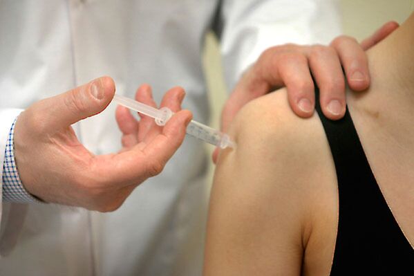
The selection of drug dosage and dosage regimen are carried out strictly individually, depending on the severity of symptoms, stage of the disease and the presence of systemic disorders. With the development of reactive synovitis, intra-articular punctures are performed with pumping out the accumulated fluid and subsequent administration of corticosteroids.
Attention:The maximum number of hormonal injections into the joint cavity is 4 times a year! Too frequent injections have a detrimental effect on the cartilage and weaken the ligamentous-tendon apparatus, which leads to "looseness" of the joint.
For severe pain accompanying severe arthrosis, opioid analgesics can be prescribed. To increase the pain threshold, drugs are usually used that are dispensed from pharmacies strictly according to a doctor’s prescription.
Chondroprotectors
Restoring cartilage tissue and slowing down its further destruction is the main goal of arthrosis therapy. Chondroprotectors successfully cope with it, but only when the disease has not gone too far. It is necessary to treat arthrosis with these remedies for several months and sometimes years.
The active ingredients of chondroprotectors are chondroitin sulfate and glucosamine, which are analogues of the structural elements of cartilage tissue. To stop the destructive process, prevent inflammation and activate the production of hyaluronic acid, intra-articular injections are performed.
It is the injections that provide maximum effect within a short period of time. In addition, a course of therapeutic injections allows you to reduce the dosage of drugs from the NSAID group.
Hyaluronic acid is part of the synovial fluid and is responsible for its viscosity, which allows bones to glide smoothly during movements. With osteoarthritis, the concentration of hyaluron in the joint fluid is significantly reduced, so intra-articular injections with hyaluronic acid are prescribed.
Local remedies
In the complex therapy of arthrosis, local agents are widely used, which can speed up recovery and prevent exacerbation. Today in pharmacies there are many different drugs that help get rid of pain and inflammation. They have anti-inflammatory, analgesic, warming and chondroprotective effects.
Only a doctor can determine how and with what to treat arthrosis in a particular patient.
The above agents have a pronounced anti-inflammatory and analgesic effect. Among the products with a warming effect, ointments with bee venom, capsicum extract, levomenthol, and capsaicin can be noted. Chondroprotectors can also be prescribed in the form of ointments.

Surgery
The indication for joint surgery is the ineffectiveness of conservative techniques and total destruction of articular cartilage. It is worth noting that radical replacement of the shoulder joint is extremely rarely required, in contrast to endoprosthetics of the joints of the lower extremities.
Surgical intervention is most often performed for post-traumatic arthrosis. After a fracture, bones may not heal properly, which leads to destruction in the cartilage and changes in the shape of the bones. With a deformed head of the humerus, endoprosthetics is the only way to restore the function of the joint.
There are several types of shoulder surgery:
- riserfacing (only the cartilage is removed, in its place an artificial prosthesis is installed);
- unipolar endoprosthetics (hemiarthroplasty) - either the head of the humerus or the articular scapula is replaced with a prosthesis;
- complete joint replacement.
Arthrosis is a chronic disease that steadily progresses. However, there are a number of preventive measures to help slow down the pathological process. The main condition for successful therapy is a gentle regime of physical activity. This does not mean a complete cessation of movement, but prolonged and intense strength exercises are absolutely contraindicated.
If you need to do physical work, you need to first stretch the joint by making several circular movements with your shoulders. And only then lift or carry something heavy. During periods of exacerbation, it is better to abandon such experiments altogether. Particular attention should be paid to any shoulder injuries, promptly consult a doctor and undergo treatment. Be healthy!